At Optimum Oral Surgery & Dental Implants, we are dedicated to bringing every smile to life. We’ve researched technology around the world and established our very own dental lab to serve our patients in New Jersey.
Our dental lab gives us the ability to reduce patients’ treatment time and provide truly exceptional experiences to both our restorative partners in the dental community and patients.
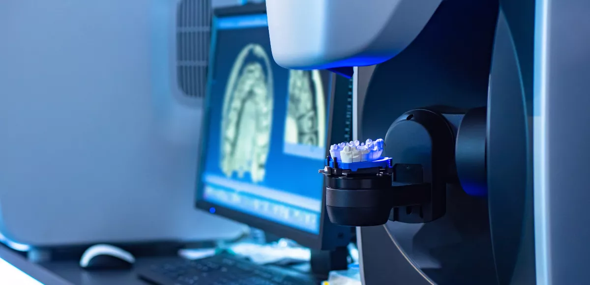
Our lab includes a 3D printer that can print a set of teeth in 20 minutes. This allows us to perform same-day tooth restoration without outsourcing to another lab. We also have a mill that customizes every set of teeth to fit the patient’s specifications.
Inside the lab, our team of dental experts use their artistic and restorative skills to finalize the look of the replacement teeth. This ensures every prosthesis is given a human touch for natural-looking results.
Benefits Of Our Own Dental Lab
Patients benefit from the convenience and technology of our dental lab. The technology allows us to provide better clinical results and enhance the patient experience:
- Our entire digital process ensures information is instantly stored and readily available
- Extreme accuracy reduces the chance of surprises during surgery for predictable results
- In-house operations allow us to provide same-day dental implants and full-arch restoration services
- The number of appointments is reduced, which saves the patient time and money
- We have complete control over the superior quality of everything our lab creates
Other Technology Used
We strive to give our patients the finest dental care possible. Our investment in advanced technology is a long-term investment in your oral health. We understand that using updated technology leads to more informed decision-making, allowing our patients to make the best possible decisions about their oral health.
Our surgical center features the latest state-of-the art technology, including:
Cone Beam Ct Scan
This technology is important to us and helps us accurately diagnose and monitor dental conditions. When facial x-rays do not provide the images that we need, we do not stop there and just make the best of it.
Instead, we will use dental cone beam computed tomography (CT). Dental Cone Beam CT is a specialized x-ray that provides clear, three-dimensional images of all pertinent structures within a patient’s mouth. We can get a closer look at the teeth of the patient, soft and hard tissues, nerve pathways, and even bone condition.
We use a dental cone beam CT to better diagnose cases of temporomandibular joint disorder (TMJ) as well as to accurately assess the condition of the nasal cavity, sinuses, nerve canals and the jaws of our patients. When our patients are experiencing pain, we sometimes use dental cone beam CT to help us determine the exact area where the pain is coming from.
The dental cone beam CT is also vital in determining treatment options for our patients with more complex dental problems. For instance, the 3D images from the dental cone beam CT can help us see where we should set dental implants and how we should address impacted teeth during surgery.
This type of advanced imaging technology is also useful in determining the tooth orientation and bone structure of the patient, as well as locating the exact position of jaw tumors. Dental cone beam CT is also a valuable tool when we need to carry out reconstructive surgeries.
X-Guide Dynamic 3D Navigation

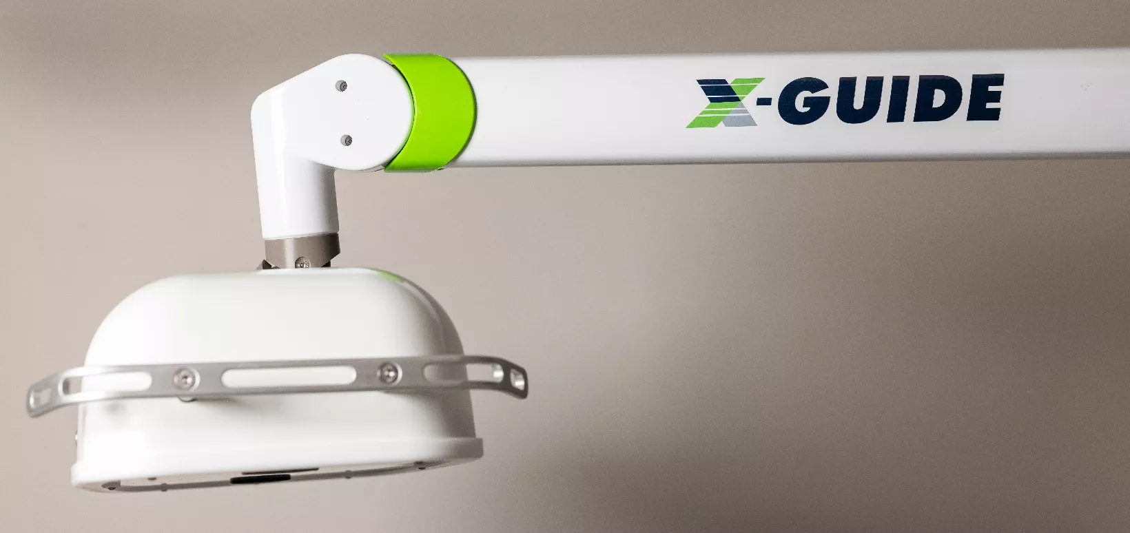
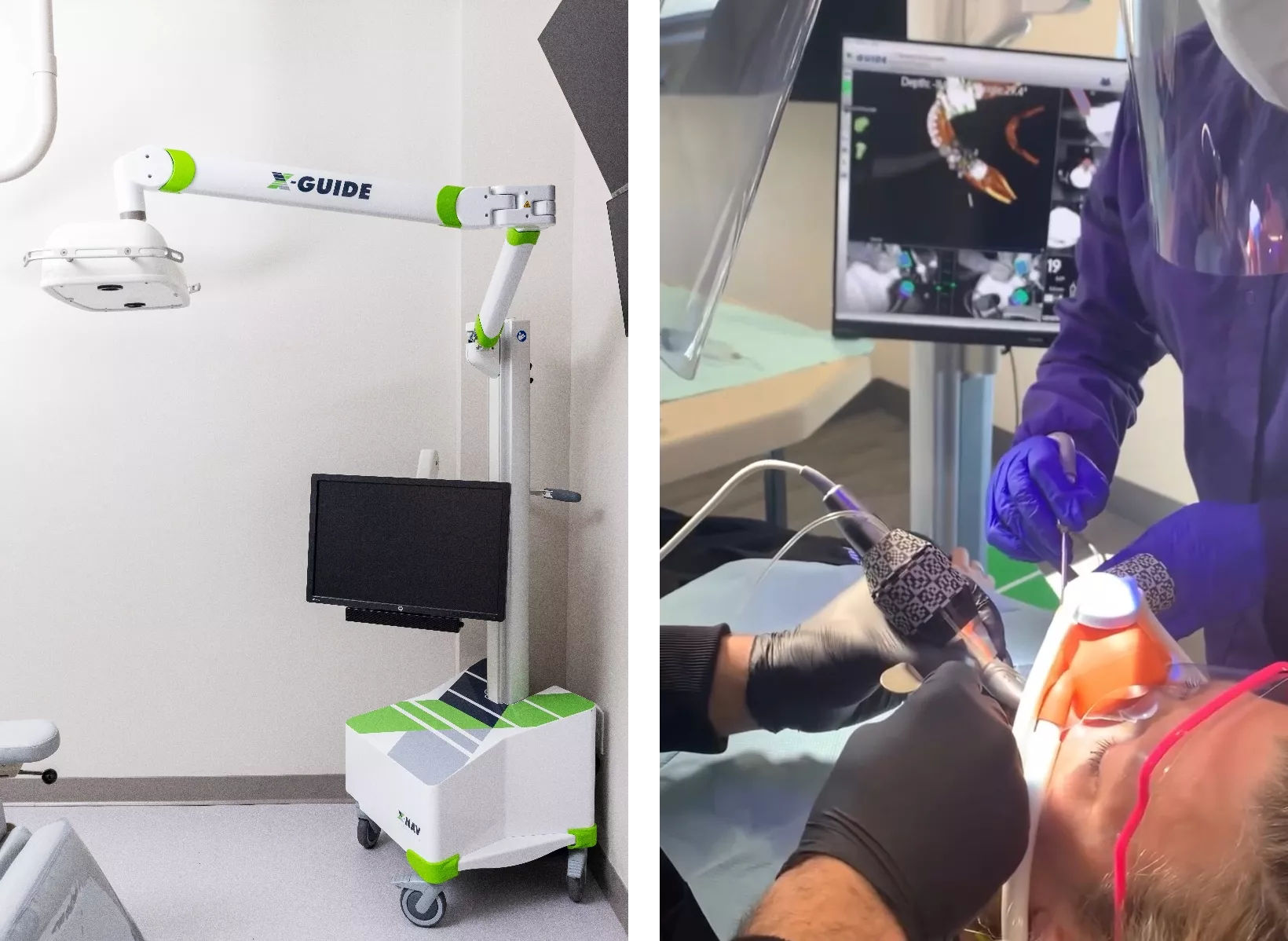
X-Guide is the newest and most advanced system approved for the placement of dental implants. This system is a valuable tool for dental professionals to improve their implant accuracy and control.
It provides real-time 3D navigation during implant surgery which allows for better visualization of the implant site and more precise placement of the implant. By integrating Cone Beam 3D imaging at Optimum Oral Surgery & Dental Implants, we can provide accuracy and precision in planning our patients’ implants and give them the highest level of care.
We can provide you with unparalleled accuracy in the placement of your dental implants and your restorations.
Thanks to 3D guided implant surgery, we can greatly reduce your risk of complications by predetermining the optimal location for your implants as well as their angle and depth.

Digital X-Rays
For decades, x-rays were taken using radiation. This fact has caused concern for many dental patients, leading them to avoid seeking necessary treatment. By avoiding treatment, the risk of severe consequences greatly increases. The risks of needing more invasive, more expensive treatments increase as well.
Along with the use of radiation, images taken with traditional x-ray technology need to be developed in a dark room. This requires taking time from your appointment to leave the room and process the images. This results in a shorter appointment with less time to go over your diagnosis and treatment options, which could then leave you confused and unsure.
Digital x-rays are a technological improvement upon traditional x-rays. Instead of radiation to capture images, digital x-rays use electronics. We place an electronic sensor inside your mouth. The sensor is attached to a computer. With the click of a button, we capture the necessary images, which are then displayed almost instantly on our computer monitor, ready for reading. There is no need to develop images or even leave the room.
Platelet Rich Fibrin (Prf)
No one wants a dry socket. It is an unwanted risk of the healing process. One of the best-known ways to prevent a dry socket is platelet rich fibrin (PRF).
In the normal healing process, your immune system sends white blood cells and growth factors to the surgery site.
These good healing cells also bring pain, inflammation, and swelling with them. PRF is a collection of those good cells, which gives you a three-day jump start on the healing process, without waiting for the swelling and inflammation to bring the good cells.
PRF reduces pain, speeds healing, and lowers the chance of getting a dry socket. PRF has many uses in medicine: for tendinitis, arthritis, plastic surgery, and in oral surgery. Using platelet rich fibrin is especially useful for those with diabetes and other conditions which impair healing due to reduced blood flow.
PRF is made from the patient’s own blood. Blood is drawn from the vein into test tubes. This blood is spun in a centrifuge while your teeth are being extracted. By the time the teeth are out, the PRF is ready. While spinning in the centrifuge, the blood separates into three layers: red blood cells, platelet rich fibrin (PRF), and watery platelet poor plasma.
The layer of PRF is yellow and gummy. It contains white blood cells, growth factors, fibrinogen (that helps with clotting), and more—all the good stuff. This gummy layer of PRF is removed from the test tube and placed in the extraction socket.
Contact Optimum Surgery Today
At Optimum Oral Surgery & Dental Implants, we are committed to providing our patients with the most advanced oral surgery care possible. That’s why we invest heavily in the latest technology and equipment.
We understand that oral surgery can be a daunting experience for some patients. That’s why we are committed to providing you with the most comfortable and stress-free experience possible. Our team of experienced oral surgeons and dental professionals will be there to guide you every step of the way.
If you have any questions about the advanced technology we use at Optimum Oral Surgery & Dental Implants, please don’t hesitate to contact us. We are here to help you achieve your oral health goals.

